
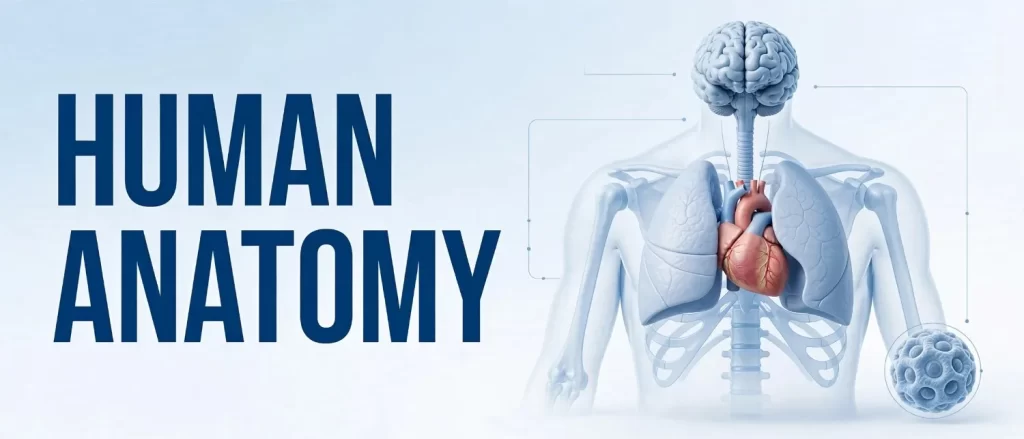
Human anatomy illustration is the specialized discipline of creating scientifically accurate visual representations of the human body’s structural organization, from macroscopic organ systems to microscopic cellular architectures. Medical professionals, educators, and publishers rely on these precise visual assets to communicate complex physiological concepts, support diagnostic reasoning, and enhance patient comprehension across clinical and academic settings. This comprehensive guide examines the methodologies, applications, and best practices that define professional-grade anatomy illustration in contemporary healthcare communication.
The visual communication of anatomical knowledge has remained a cornerstone of medical science since the Renaissance era. Today, human anatomy illustration serves as an indispensable bridge between raw clinical data and actionable medical understanding. Whether you are a medical educator designing curriculum materials, a surgeon preparing for a complex procedure, or a publisher assembling a peer-reviewed journal, the quality of your visual assets directly impacts knowledge retention and clinical outcomes.
If your organization requires medical illustration services that combine scientific rigor with artistic precision, partnering with a specialized studio ensures your visual communication meets the highest standards of accuracy and clarity.
What is Human Anatomy Illustration?
Human anatomy illustration encompasses the creation of detailed visual depictions that accurately represent the form, position, structure, and relationships of human body components. Unlike general artistic rendering, this discipline demands an intimate understanding of gross anatomy, histology, embryology, and pathophysiology. Every line, shade, and color choice must align with established medical knowledge and current anatomical nomenclature.

The scope of human anatomy illustration extends far beyond textbook diagrams. Modern practitioners produce 3D medical illustrations for surgical planning, interactive digital models for virtual dissection labs, and vector-based graphics for regulatory submissions. The unifying thread across all these applications is an unwavering commitment to anatomical fidelity and educational effectiveness.
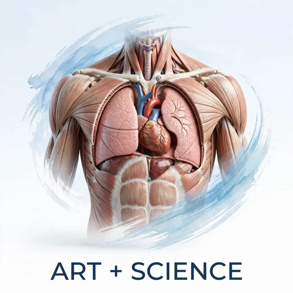
The Intersection of Art and Science
At its core, human anatomy illustration exists at the intersection of artistic skill and biomedical expertise. A professional medical illustrator typically holds advanced training in both fine arts and human anatomy, often completing graduate-level certification from accredited programs. This dual competency enables them to translate dense clinical information into visually intuitive formats without sacrificing scientific accuracy.
The artistic dimension involves mastery of perspective, lighting, texture, and composition. The scientific dimension requires fluency in anatomical terminology, physiological processes, and disease pathology. When these competencies converge, the result is visual content that educates, informs, and engages diverse audiences ranging from first-year medical students to seasoned specialists.
The Evolution of Anatomy Illustration
The history of anatomical visualization reflects broader shifts in medical knowledge and technological capability. Understanding this evolution provides valuable context for appreciating the sophistication of contemporary illustration practices.
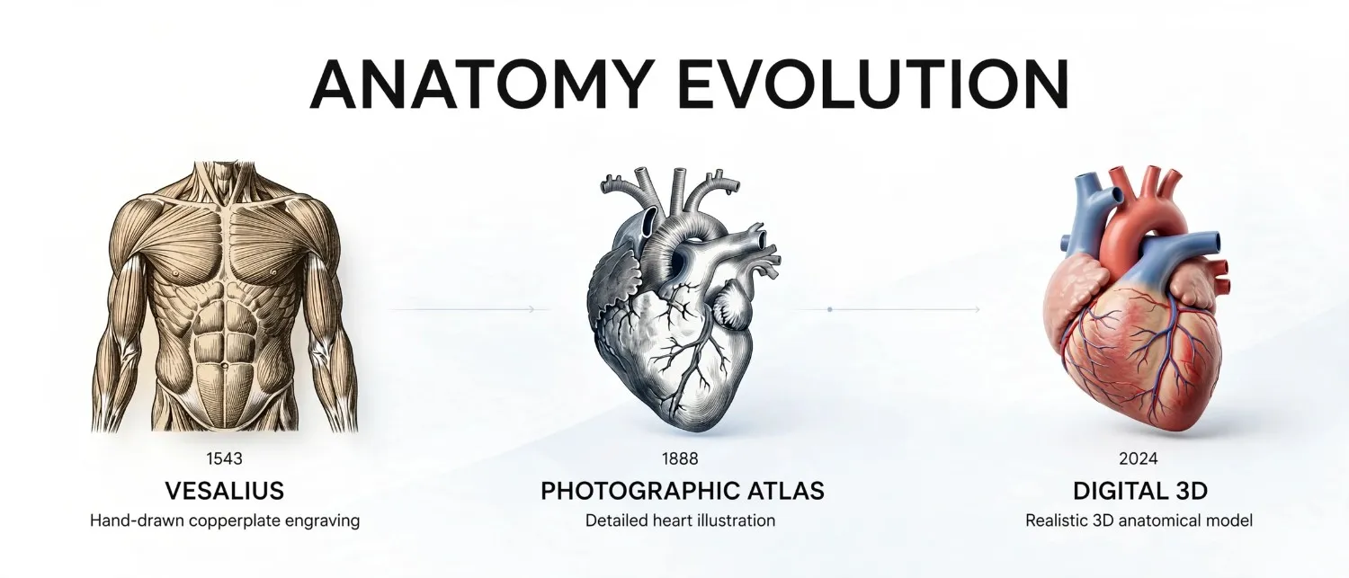
From Vesalius to Digital Rendering
Andreas Vesalius revolutionized anatomical communication in 1543 with De Humani Corporis Fabrica, establishing standards for anatomical accuracy that persist today. Hand-drawn copperplate engravings dominated medical publishing for centuries, offering unparalleled detail but limited reproducibility. The twentieth century introduced photographic atlases, which provided realism but often lacked the clarity of idealized illustrations.
The digital revolution transformed the field entirely. Computer-generated imagery, vector graphics, and real-time 3D modeling now enable illustrators to depict structures from impossible angles, animate physiological processes, and customize visuals for specific educational objectives. This technological progression has expanded the utility of human anatomy illustration across virtually every domain of healthcare communication.
The Impact of 3D Technology
Three-dimensional visualization represents perhaps the most significant advancement in anatomical illustration since the invention of the printing press. Contemporary 3D medical illustration software allows artists to construct fully manipulable models of organs, vascular networks, and skeletal structures. These assets support immersive learning experiences, preoperative surgical simulation, and patient education initiatives that were impossible with traditional two-dimensional media.
The transition from static images to interactive 3D environments reflects a broader pedagogical shift toward experiential learning. Medical students who manipulate virtual anatomical models demonstrate improved spatial reasoning and retention compared to those relying solely on textbook diagrams. Clinicians use 3D reconstructions from patient imaging data to plan interventions with unprecedented precision.
Types of Human Anatomy Illustrations
Medical professionals encounter diverse illustration formats, each optimized for specific communication objectives. Understanding these categories helps stakeholders select appropriate visual strategies for their particular needs.
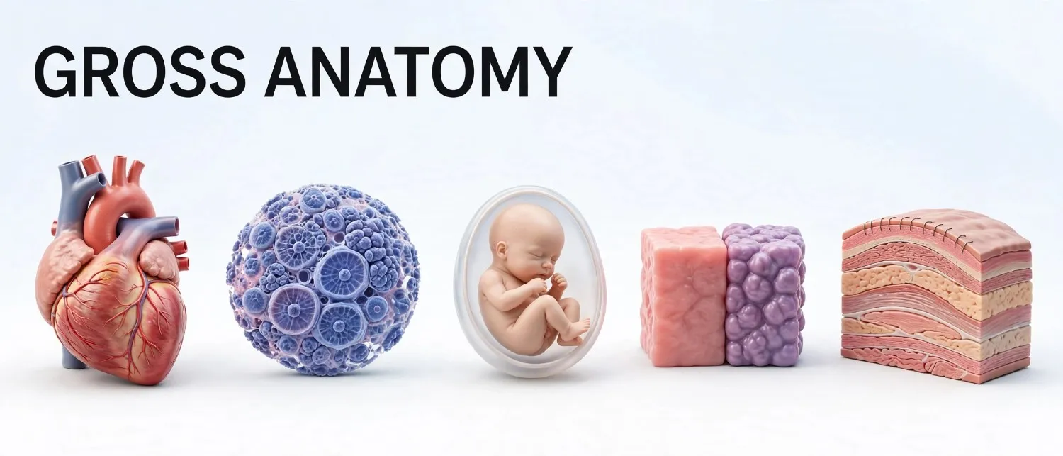
Gross Anatomy Illustrations
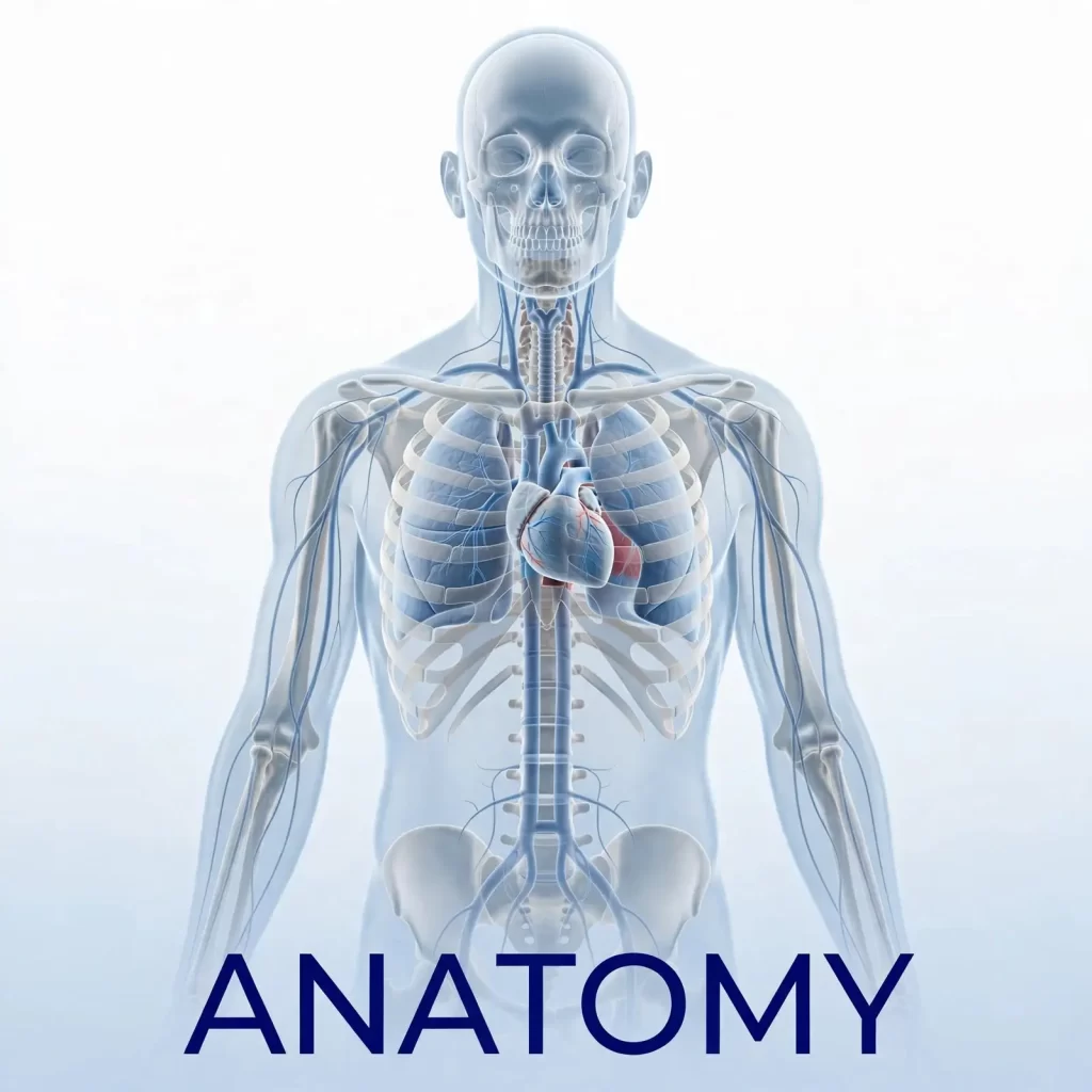
Gross anatomy illustrations depict structures visible to the naked eye, including organ systems, muscle groups, skeletal elements, and vascular networks. These visuals typically emphasize spatial relationships, topographical landmarks, and functional correlations. High-quality gross anatomy illustrations simplify complex three-dimensional relationships into comprehensible two-dimensional representations without distorting critical anatomical truths.
Microscopic and Histological Illustrations
Histological illustrations translate cellular and tissue-level architecture into accessible visual formats. These images support pathology education, research communication, and diagnostic training. Unlike photomicrographs, which capture actual specimens with all their variability and artifacts, illustrated histological images present idealized representations that highlight diagnostically relevant features while minimizing distracting elements.
Developmental and Embryological Illustrations
Embryological illustrations trace anatomical development from fertilization through fetal maturation. These visuals are particularly valuable for explaining congenital anomalies, developmental timelines, and the morphological transformations underlying organogenesis. Given the dynamic and often abstract nature of embryological processes, illustration frequently offers clearer communication than photographic documentation.
Pathological and Surgical Illustrations
Pathological illustrations depict disease states, traumatic injuries, and post-surgical anatomy with clinical precision. These images support case documentation, informed consent discussions, and medical-legal communication. Surgical illustrations, in particular, require intimate familiarity with operative techniques and postoperative anatomy to accurately represent procedural steps and expected outcomes.
Comparative and Evolutionary Illustrations
Comparative anatomy illustrations juxtapose human structures with homologous elements in other species, supporting research communication and veterinary crossover education. Evolutionary illustrations trace anatomical adaptations across phylogenetic lineages, providing context for understanding human morphological peculiarities.
Applications in Medical Education

The educational value of human anatomy illustration cannot be overstated. Visual learning constitutes a primary modality for most medical students, and well-designed illustrations significantly enhance comprehension, retention, and recall.
Supporting Visual-Spatial Learning
Medical education demands sophisticated spatial reasoning capabilities. Students must mentally reconstruct three-dimensional anatomy from two-dimensional cross-sections, anticipate structural variations, and visualize operative approaches. High-quality anatomical illustrations scaffold this cognitive development by providing clear, consistent reference images that emphasize relevant spatial relationships.
Research consistently demonstrates that students who study with professionally illustrated materials outperform peers relying solely on text or uncurated photographic resources. The idealization inherent in illustration eliminates irrelevant visual noise, directing attention toward educationally salient features.
Standardizing Educational Content
Anatomical variation presents a genuine challenge for medical educators. Real specimens and photographs display normal variation, pathological changes, and postmortem artifacts that can confuse novice learners. Illustrations offer the advantage of presenting standardized, idealized anatomy that establishes baseline knowledge before students encounter biological variability.
This standardization proves particularly valuable in distance learning environments, where students may lack access to cadaveric specimens or physical models. Digital illustration libraries enable consistent, high-quality anatomical instruction across geographically distributed learner populations.
Enhancing Assessment Materials
Examination items in medical education frequently incorporate anatomical illustrations to assess spatial reasoning and anatomical knowledge. Well-designed illustrations ensure that assessment focuses on conceptual understanding rather than interpretation of poor-quality images. Professional illustration also supports the creation of question banks, virtual patient cases, and adaptive learning modules.
Applications in Clinical Practice

Beyond education, human anatomy illustration serves numerous practical functions in direct patient care and clinical operations.
Surgical Planning and Navigation
Surgeons increasingly rely on illustrated anatomical maps derived from preoperative imaging. These visualizations clarify patient-specific anatomy, identify critical structures to preserve, and communicate operative plans to surgical teams. In complex cases involving variant anatomy or revision surgery, custom illustrations provide clarity that standard imaging alone cannot achieve.
Patient Education and Informed Consent
Effective patient communication requires translating medical complexity into accessible language and visuals. Anatomical illustrations help patients understand their diagnoses, appreciate proposed interventions, and provide truly informed consent. Research indicates that patients who receive visual explanations demonstrate higher comprehension, lower anxiety, and greater treatment adherence.
Custom illustrations tailored to individual patient anatomy offer particular value in informed consent discussions. Generic diagrams may fail to capture patient-specific pathology or anatomical variants, whereas personalized visuals ensure that discussions address relevant clinical realities.
Medical Device and Pharmaceutical Communication
The medical device industry depends on anatomical illustration to demonstrate product positioning, mechanism of action, and proper usage. Regulatory submissions, instruction manuals, and marketing materials all require accurate anatomical visuals that satisfy both scientific and commercial communication standards. Similarly, pharmaceutical companies use anatomical illustration to explain drug mechanisms, target tissues, and physiological effects.
Applications in Scientific Publishing
Peer-reviewed medical literature demands visual content that meets rigorous standards of accuracy, clarity, and reproducibility. Human anatomy illustration plays a vital role in communicating research findings across diverse publication formats.

Journal Articles and Review Papers
Medical and scientific journals routinely publish anatomical illustrations to clarify methodology, present novel findings, and summarize complex systems. These images must satisfy both aesthetic and scientific review, often undergoing scrutiny by subject matter experts, editors, and peer reviewers. Understanding author guidelines for illustrations in medical scientific journals is essential for researchers seeking publication success.
High-impact journals increasingly favor original illustrations over stock imagery or unmodified photographs. Original artwork ensures that visuals precisely match the article’s content, eliminating the mismatches and inaccuracies that can undermine credibility.
Textbook and Atlas Production
Medical textbooks and anatomical atlases represent the most traditional and enduring application of professional anatomy illustration. These publications require extensive image libraries covering comprehensive anatomical content. Contemporary atlas production increasingly incorporates digital supplements, interactive features, and layered illustrations that allow readers to progressively reveal deeper anatomical structures.
Research Communication and Grant Proposals
Visual content significantly impacts the persuasiveness of research proposals and conference presentations. Well-designed anatomical illustrations help reviewers grasp complex hypotheses, appreciate methodological innovations, and recognize the significance of proposed investigations. In competitive funding environments, professional visual communication can distinguish successful proposals from unsuccessful competitors.
For researchers preparing publication-quality visuals, exploring scientific illustration images for research papers provides valuable guidance on standards and best practices.
The Human Anatomy Illustration Process
Creating professional anatomical illustrations involves a structured workflow that ensures scientific accuracy, visual clarity, and alignment with client objectives. Understanding this process helps medical professionals collaborate effectively with illustrators and articulate their visualization needs.
Step 1: Research and Reference Gathering

Every illustration project begins with comprehensive research. The illustrator reviews relevant anatomical literature, examines imaging studies, and consults subject matter experts to establish a thorough understanding of the content. For custom projects, this phase may involve reviewing patient-specific imaging or proprietary research data.
Reference materials typically include anatomical atlases, peer-reviewed articles, surgical photographs, and diagnostic imaging. The illustrator evaluates these sources for accuracy, currency, and relevance, identifying any inconsistencies or gaps requiring clarification.
Step 2: Concept Development and Sketching

With research foundations established, the illustrator develops preliminary sketches exploring composition, perspective, and emphasis. These rough drafts present structural relationships and narrative flow without investing in detailed rendering. Client feedback at this stage ensures that the illustration direction aligns with communication objectives before significant production time is committed.
Concept development considers the intended audience, publication format, and viewing context. An illustration designed for a surgical atlas requires different compositional choices than one created for a patient education brochure.
Step 3: Detailed Rendering and Refinement

Approved sketches progress to detailed rendering, where anatomical structures receive precise delineation, appropriate shading, and accurate coloration. Digital tools enable non-destructive editing, layering, and iterative refinement throughout this phase. The illustrator continually cross-references anatomical sources to verify accuracy.
Rendering quality depends heavily on technical proficiency with illustration software, understanding of anatomical form, and sensitivity to visual hierarchy. Effective illustrations direct viewer attention toward primary structures while maintaining contextual accuracy in surrounding anatomy.
Step 4: Scientific Review and Revision

Completed illustrations undergo expert review by medical professionals, researchers, or subject matter specialists. Reviewers evaluate anatomical accuracy, clinical relevance, and educational effectiveness. Constructive feedback drives revision cycles that refine the illustration until it satisfies all stakeholders.
This review process is non-negotiable for professional medical illustration. Even experienced illustrators benefit from expert validation, particularly when depicting rare anatomical variants, novel surgical techniques, or emerging therapeutic interventions.
Step 5: Final Production and Delivery

Following approval, illustrations are prepared for final delivery in formats appropriate to their intended use. Vector files suit publication and scaling requirements, while raster formats accommodate web and presentation needs. Metadata, color profiles, and resolution specifications are optimized for the target output.
Common Mistakes in Anatomy Illustration
Even well-intentioned anatomical visualization efforts can falter when common pitfalls are not anticipated and avoided.
Prioritizing Aesthetics Over Accuracy
The most frequent error in anatomical illustration involves sacrificing scientific precision for visual appeal. While aesthetic quality matters, it must never compromise anatomical truth. Structures must maintain correct proportions, positions, and relationships even when doing so complicates composition or reduces dramatic impact.
Overloading Single Images
Attempting to communicate excessive information within a single illustration produces visual clutter and cognitive overload. Effective anatomical communication distributes information across multiple images or progressive disclosure formats. Each illustration should have a clear, singular focus that guides viewer attention.
Neglecting Audience Appropriateness
Illustrations designed for specialist surgeons may overwhelm medical students, while simplified patient education diagrams may frustrate researchers seeking granular detail. Audience analysis must inform every illustration decision, from level of detail to terminology integration.
Ignoring Contextual Anatomy
Isolating target structures without adequate contextual anatomy can mislead viewers regarding spatial relationships and topographical orientation. Effective illustrations provide sufficient surrounding structure to orient viewers while maintaining emphasis on primary subjects.
Using Outdated Anatomical Knowledge
Medical understanding evolves continuously. Illustrations based on outdated terminology, disproven concepts, or superseded anatomical models undermine credibility and educational value. Professional illustrators maintain currency with evolving anatomical knowledge and nomenclature standards.
Expert Tips for Medical Professionals

Medical professionals who collaborate with illustrators or commission custom anatomical visuals can maximize project outcomes by following established best practices.
Tip 1: Define Clear Communication Objectives
Before engaging an illustrator, articulate precisely what the illustration must communicate, to whom, and in what context. Specific objectives regarding educational level, clinical application, and publication format guide illustration decisions and prevent costly revisions.
Tip 2: Provide Comprehensive Reference Materials
The quality of reference materials directly impacts illustration accuracy. Provide illustrators with relevant imaging studies, literature citations, and expert contacts. For patient-specific illustrations, de-identified diagnostic images and operative reports offer invaluable guidance.
Tip 3: Engage Early and Iterate Often
Involving illustrators at project inception rather than as afterthoughts yields superior results. Early engagement allows illustrators to contribute to communication strategy, suggest optimal visual approaches, and identify potential challenges before they become costly problems.
Tip 4: Respect Professional Expertise
Medical professionals possess deep clinical knowledge, but illustrators bring specialized expertise in visual communication. Trust their judgment regarding composition, color, and format decisions that may differ from initial expectations but serve communication objectives more effectively.
Tip 5: Plan for Multi-Platform Usage
Contemporary medical communication spans print, digital, presentation, and mobile platforms. Discussing multi-platform requirements early ensures illustrations are created in versatile formats that maintain quality across diverse outputs.
Comparison of Anatomy Illustration Formats
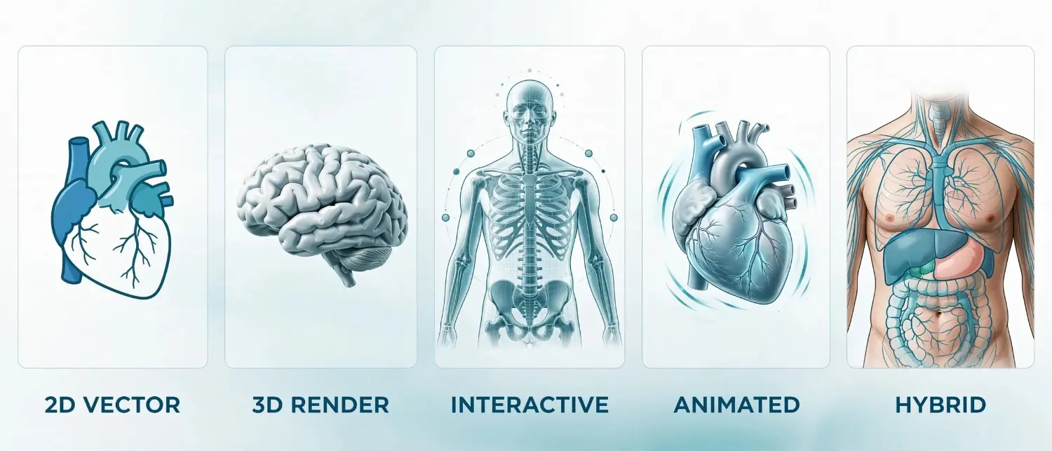
Selecting the appropriate illustration format depends on communication objectives, audience characteristics, and technical constraints. The following comparison table summarizes key distinctions among common formats.
| Format | Best For | Advantages | Limitations |
|---|---|---|---|
| 2D Vector Illustration | Textbooks, journals, presentations | Infinitely scalable, clean lines, easy to edit | Limited depth perception, static viewing angle |
| 3D Rendered Illustration | Surgical planning, patient education | Realistic depth, multiple viewing angles possible | Higher production time, specialized software required |
| Interactive Digital Model | E-learning, virtual dissection | User manipulation, layered disclosure | Technical complexity, platform compatibility issues |
| Animated Illustration | Physiology education, mechanism of action | Depicts dynamic processes, high engagement | Longer production timeline, larger file sizes |
| Hybrid Photo-Illustration | Clinical case documentation | Combines realism with clarity | Requires quality source photography |
Essential Tools and Technologies
The modern anatomy illustrator’s toolkit spans traditional artistic media and cutting-edge digital platforms. Understanding available technologies helps clients appreciate production capabilities and constraints.
Digital Illustration Software
Industry-standard applications include Adobe Illustrator for vector work, Adobe Photoshop for raster rendering, and specialized medical illustration platforms. 3D medical illustration software such as ZBrush, Maya, and Blender enable sophisticated three-dimensional modeling and rendering. Proprietary medical visualization platforms offer anatomically accurate base models that illustrators customize for specific projects.
Anatomical Reference Databases
Professional illustrators maintain extensive reference libraries including anatomical atlases, imaging archives, and specimen photography. Digital resources such as the National Library of Medicine’s Visible Human Project and academic anatomical databases provide invaluable reference material for accurate illustration.
Hardware Considerations
High-resolution displays, graphics tablets, and calibrated monitors ensure color accuracy and detail visibility throughout the production process. For 3D illustration, powerful workstations with dedicated graphics processing capabilities accelerate rendering and enable real-time model manipulation.
The Future of Human Anatomy Illustration
Emerging technologies promise to reshape how medical professionals create, consume, and interact with anatomical visualizations. Anticipating these developments positions organizations to leverage new capabilities effectively.

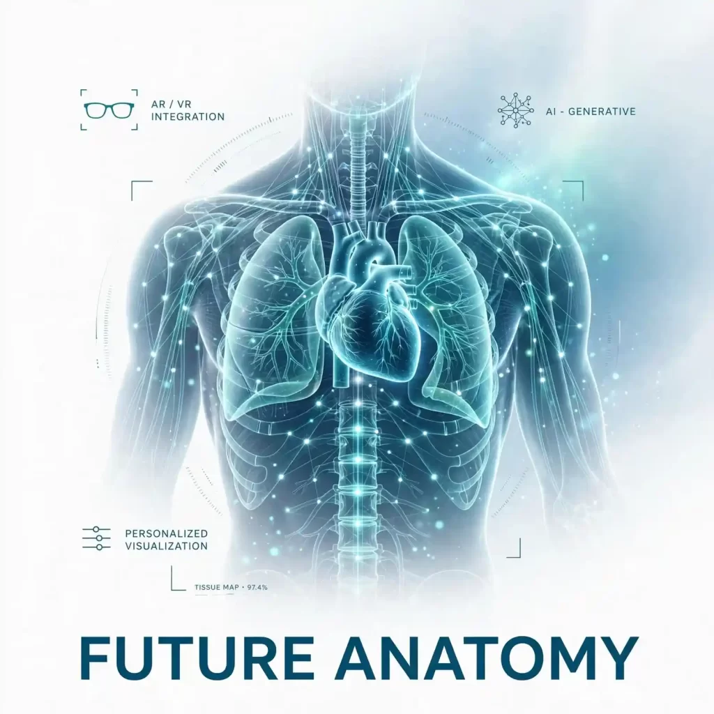
Artificial Intelligence and Generative Tools
AI-assisted illustration tools increasingly support anatomical visualization workflows, automating routine tasks and accelerating production timelines. However, human expertise remains essential for ensuring scientific accuracy, clinical relevance, and educational effectiveness. The most promising applications combine AI efficiency with professional illustrator oversight.
Augmented and Virtual Reality Integration
AR and VR technologies enable immersive anatomical exploration that transcends traditional illustration limitations. Medical students can virtually dissect three-dimensional bodies, surgeons can overlay anatomical guidance onto live operative fields, and patients can visualize their own anatomy through personalized reconstructions. These modalities demand new illustration approaches that account for spatial interaction and real-time rendering constraints.
Personalized Anatomical Visualization
Advances in medical imaging and computational modeling enable increasingly personalized anatomical visualization. Patient-specific illustrations derived from individual CT, MRI, or ultrasound data support precision medicine initiatives, customized surgical planning, and tailored patient education. This trend toward personalization demands illustrators who can integrate clinical data with artistic communication.
The distinction between medical illustration and medical animation continues to blur as interactive and motion-based formats proliferate. Modern medical communication increasingly requires hybrid skill sets that span static, animated, and interactive visualization.
Frequently Asked Questions
What is human anatomy illustration?
Human anatomy illustration is the specialized practice of creating scientifically accurate visual representations of human body structures for educational, clinical, and research purposes. These illustrations range from macroscopic organ system diagrams to microscopic cellular depictions, all designed to communicate anatomical relationships with precision and clarity.
How does human anatomy illustration differ from medical photography?
While medical photography captures actual specimens or clinical presentations, human anatomy illustration creates idealized representations that emphasize educationally relevant features while eliminating distracting artifacts, variations, or pathological changes. Illustrations offer standardization, clarity, and the ability to depict structures from optimal viewing angles impossible to achieve with photography alone.
What qualifications should a professional medical illustrator possess?
Professional medical illustrators typically hold graduate degrees from accredited medical illustration programs, combining advanced training in human anatomy, physiology, and pathology with artistic development in drawing, digital rendering, and visual design. Many maintain certification through professional organizations and engage in continuing education to remain current with evolving medical knowledge.
How long does it take to create a custom anatomy illustration?
Production timelines vary based on complexity, research requirements, and revision cycles. Simple diagrams may require several days, while complex 3D renderings or extensive anatomical atlases can require weeks or months. Early engagement with clear objectives and comprehensive reference materials significantly accelerates the process.
What file formats are best for medical anatomy illustrations?
Vector formats such as AI, EPS, and SVG suit publication and scaling needs, while high-resolution raster formats like TIFF and PNG accommodate web and presentation requirements. For 3D assets, OBJ, FBX, and proprietary format deliverables depend on intended usage platforms. Discussing format requirements during project initiation ensures appropriate deliverables.
Can anatomy illustrations be used for patient-specific surgical planning?
Yes, custom anatomy illustrations derived from patient imaging data increasingly support surgical planning, informed consent, and interdisciplinary communication. These personalized visuals clarify patient-specific anatomy, identify critical structures, and help surgical teams anticipate operative challenges.
How do I ensure anatomical accuracy in published illustrations?
Engage qualified medical illustrators with appropriate training, provide comprehensive reference materials, and implement expert review processes involving subject matter specialists. Cross-referencing illustrations against authoritative anatomical sources and current nomenclature standards further ensures accuracy.
Conclusion
Human anatomy illustration remains an essential discipline within modern healthcare communication, bridging the gap between complex biomedical knowledge and accessible visual understanding. From medical education and clinical practice to scientific publishing and patient engagement, professionally crafted anatomical visuals enhance comprehension, support decision-making, and elevate the quality of medical communication.

The evolution from hand-drawn atlases to interactive 3D environments reflects both technological advancement and persistent demand for accurate, engaging anatomical visualization. As medical knowledge expands and communication technologies evolve, the role of skilled anatomical illustration will only grow in importance.
Organizations seeking to distinguish their educational materials, clinical communications, or research publications should invest in professional illustration services that deliver scientific precision alongside visual excellence. The return on this investment manifests in improved learner outcomes, enhanced patient understanding, and stronger professional credibility.
Ready to elevate your medical communication with precision-crafted anatomical visuals? Contact our team today to discuss your human anatomy illustration needs and discover how specialized visual expertise can transform your educational, clinical, or publishing objectives into compelling, scientifically rigorous visual content.