
The history of medical illustration traces the evolution of anatomical visualization from Galenic anatomical texts and hand-copied manuscripts to Andreas Vesalius’s 1543 De humani corporis fabrica, Max Brödel’s carbon dust technique, and today’s DICOM-driven 3D rendering. Each stage replaced inference with direct observation, which shifted anatomy education from textual authority to reproducible, evidence-based imagery. Modern practice now combines surgical-grade accuracy, volumetric segmentation, and digital publishing workflows to deliver scalable visual communication across journals, clinics, and device marketing.
Whether you publish research, market a device, or teach clinicians, this lineage explains why accuracy matters so much in visual work. If you need anatomically accurate 3D medical visualization services today, the lessons below show what separates reliable imagery from simple decoration.
Quick Answer: What Is the History of Medical Illustration?
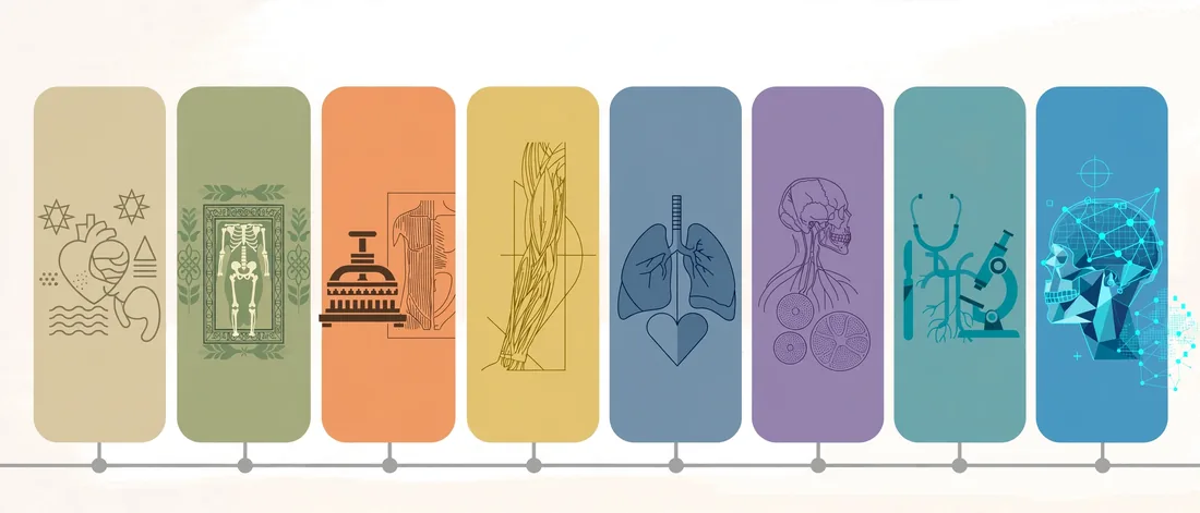
The history of medical illustration is the story of how anatomical and surgical knowledge became visible, shareable, and verifiable. It started with simple diagrams in ancient and medieval texts. Then it accelerated during the Renaissance, when artists began drawing from dissected bodies. Today it relies on 3D modeling, scan data, and interactive media.
Here are the seven milestones that define the field:
- Ancient world (c. 1600 BCE – 200 CE): Egyptian, Greek, and Indian texts describe anatomy and surgery, mostly in words rather than images.
- 1316 – 1491: Mondino de Luzzi’s dissection text and the Fasciculus medicinae bring anatomical images into medical education.
- c. 1489 – 1515: Leonardo da Vinci records human anatomy through direct observation.
- 1543: Andreas Vesalius publishes the Fabrica and sets a new standard for accuracy.
- 1747 – 1858: Albinus, William Hunter, and Henry Gray refine precision through engraving and woodcut.
- 1911: Max Brödel founds the first formal department of medical art at Johns Hopkins.
- 1990s onward: Digital imaging, the Visible Human Project, and 3D software transform the workflow.
For a wider primer on the discipline itself, read our complete overview of what medical illustrators create and why.
Timeline: The History of Medical Illustration at a Glance
This table gives you a fast, scannable view of the major eras. Use it as a reference as you read the detailed sections below.
| Era | Years | Key Figure or Work | Main Medium | Why It Mattered |
|---|---|---|---|---|
| Ancient | c. 1600 BCE – 200 CE | Edwin Smith Papyrus, Herophilus, Galen | Papyrus, text, sketches | Established early surgical and anatomical knowledge |
| Medieval | 500 – 1450 | Mondino de Luzzi, illuminated manuscripts | Hand-painted codices | Preserved and standardized anatomical teaching |
| Early print | 1491 – 1520s | Fasciculus medicinae | Woodcut | Made anatomical images reproducible |
| Renaissance | 1489 – 1543 | Leonardo da Vinci, Andreas Vesalius | Pen, chalk, woodcut | Put dissection-based observation first |
| Early modern | 1600 – 1800 | Albinus, Hunter, Gautier d’Agoty | Copperplate, mezzotint, wax | Added measured accuracy and color |
| 19th century | 1800 – 1900 | Bourgery, Gray, Carter | Lithography, woodcut, photography | Scaled anatomy for students and surgeons |
| Professional era | 1894 – 1960 | Max Brödel, Frank Netter | Carbon dust, watercolor, gouache | Created training programs and clinical standards |
| Digital era | 1987 – today | Visible Human Project, 3D artists | CT/MRI data, 3D software, VR | Enabled interactive, data-driven visuals |
Ancient Foundations: Anatomy Before Accurate Images
Early medicine depended on observation of injuries, rituals, and animal dissection. As a result, ancient anatomical knowledge came with real gaps. Nevertheless, these early efforts planted the seeds for everything that followed.
Egypt, India, and China: Early Written Anatomy
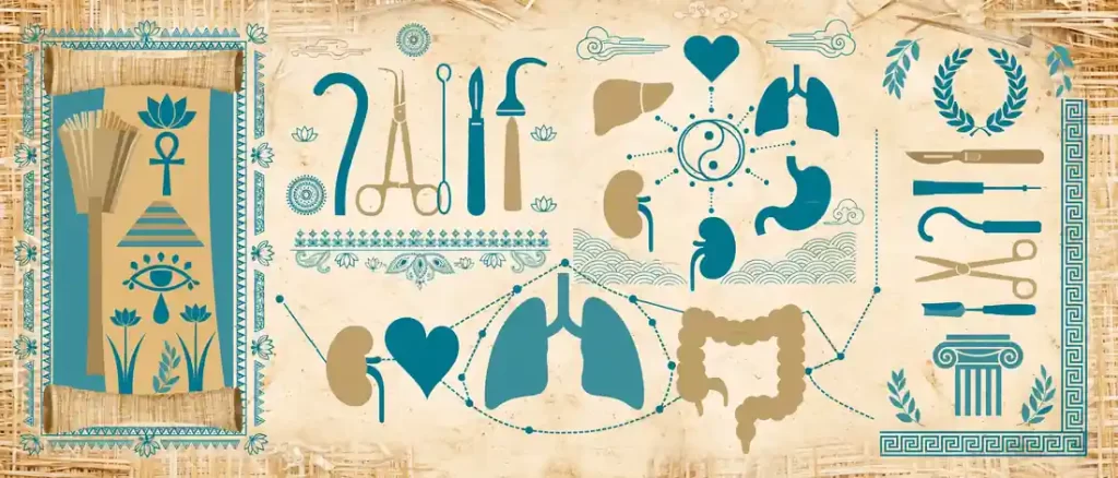
The Edwin Smith Papyrus, which dates to roughly 1600 BCE, describes dozens of trauma cases with methodical observations. Similarly, the Sushruta Samhita in ancient India documents surgical procedures and instruments in detail. Chinese medical classics also described organs and channels, although they emphasized function over structure.
These sources shared a limitation. Authors relied mostly on text, so readers had to imagine the structures. Consequently, accuracy depended on the skill of the writer and the memory of the student.
Greek Alexandria and Galen’s Long Shadow
In third-century BCE Alexandria, Herophilus and Erasistratus performed some of the earliest systematic human dissections. Later, Galen of Pergamon (129 – c. 216 CE) shaped Western medicine for well over a thousand years. However, Galen worked largely with animals such as pigs and macaques, so his descriptions contained human errors.
Those errors survived for centuries because scholars copied Galen’s texts by hand. Furthermore, few people dared to challenge an authority so revered. This is the problem that Renaissance illustrators would eventually solve.
Key takeaway: Ancient anatomy depended on words and authority. Visual proof arrived much later.
Medieval Manuscripts and the First Printed Anatomy Books
During the medieval period, monks and university scribes copied anatomical texts into illuminated manuscripts. The images were symbolic rather than realistic. For example, the “zodiac man” linked body parts to astrological signs, while the “wound man” catalogued injuries a surgeon might meet.

Still, a few milestones pushed the field forward:
- Mondino de Luzzi’s Anathomia (1316): This Bologna text promoted dissection as a teaching method and became a standard university reference.
- Persian and Arabic traditions: Scholars such as Ibn Sina preserved Greek medicine and added commentary, and later Persian manuscripts included full-body anatomical diagrams.
- The Fasciculus medicinae (1491): This Venetian volume ranks among the earliest illustrated printed medical books, and it showed a dissection scene alongside other medical subjects.
Printing changed the economics of visual knowledge. Previously, every copy required a scribe and an artist. Afterward, one woodblock could produce hundreds of identical images. Therefore, the history of medical illustration entered a new phase: reproducibility.
The Renaissance: Leonardo and the Birth of Observational Anatomy
The Renaissance valued direct observation, and anatomy benefited enormously. Artists and physicians began to share dissection rooms. Moreover, artists wanted to understand muscle and bone to paint the human form convincingly.
Leonardo da Vinci’s Anatomical Drawings
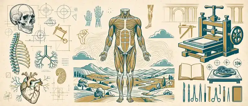
Leonardo da Vinci (1452 – 1519) filled notebooks with studies of the skull, spine, heart, and fetus between roughly 1489 and 1515. He drew structures from multiple angles, added cross-sections, and used written annotations to explain function. In many ways, he anticipated techniques that modern illustrators still use.
Unfortunately, Leonardo never published his anatomical work during his lifetime. His drawings stayed largely out of public view for centuries. Consequently, they did not reshape medical education the way Vesalius’s printed book did.
Andreas Vesalius and De Humani Corporis Fabrica (1543)
Andreas Vesalius (1514 – 1564) was only 28 when his De humani corporis fabrica libri septem appeared in Basel in 1543. The seven-book work combined careful dissection, systematic description, and large woodcut plates. According to Britannica’s biography of Vesalius, his work challenged Galen’s authority and laid the foundation for modern anatomy.
Vesalius did not work alone. Scholars generally attribute the illustrations to artists connected with Titian’s circle in Venice, and Jan Stephan van Calcar is often named, although the exact attribution remains debated. The collaboration between anatomist, artist, and printer set a template that medical illustration still follows today.
Here is what made the Fabrica revolutionary:
- Evidence-first approach: Vesalius dissected human bodies himself and corrected many of Galen’s claims.
- Integrated art and text: Images and descriptions worked together instead of sitting on separate pages.
- Expressive muscle figures: Posed bodies stood in continuous landscapes, which made structure easier to grasp.
- Companion Epitome: A shorter version served students who needed a compact reference.
- Print quality: Basel’s printing community produced detailed woodcuts at a scale that earlier books could not match.
Why the Fabrica Still Defines the History of Medical Illustration
The Fabrica proved that images could carry evidence, not just decoration. In addition, it showed that accuracy requires collaboration between experts and artists. Today’s studios follow the same logic when a surgeon reviews a rendering before publication.
Therefore, when people ask where modern medical illustration began, most scholars point to 1543. Leonardo gave the field its curiosity. Vesalius gave it a publishing model.
17th and 18th Centuries: Copperplate, Color, and Wax
After Vesalius, anatomists raced to refine his work. Meanwhile, new printing methods and instruments expanded what illustrators could show.
Engraving and the Microscope
Copperplate engraving replaced woodcut for many anatomical works because it captured finer lines and subtle shading. At the same time, microscopes opened an invisible world. Robert Hooke’s Micrographia (1665) amazed readers with detailed drawings of tiny structures, while early microscopists such as Malpighi and Leeuwenhoek documented capillaries and cells.
As a result, illustrators gained a new job. They now had to translate what only a lens could reveal.
Albinus, Hunter, and the Pursuit of Measured Accuracy
Two eighteenth-century works stand out:
- Bernhard Siegfried Albinus, Tabulae sceleti et musculorum corporis humani (1747): Illustrator Jan Wandelaar worked from a measured system that helped him reproduce proportions precisely.
- William Hunter, The Anatomy of the Human Gravid Uterus (1774): Jan van Rymsdyk’s life-sized, naturalistic drawings documented pregnancy anatomy with remarkable realism.
These books show an important shift. Accuracy became a method, not a hope. Likewise, the best illustrators worked with strict observation protocols that today’s teams still echo.
Color Printing and Anatomical Wax Models
Jacques Fabien Gautier d’Agoty pioneered color mezzotint anatomical prints from the 1740s. His multi-plate process produced vivid images, even if critics sometimes questioned their anatomical precision.
In parallel, wax modelers in Florence and Vienna built three-dimensional teaching tools. Clemente Susini’s models at La Specola and the Josephinum collection in Vienna let students study anatomy without constant access to cadavers. In effect, these were the ancestors of today’s 3D anatomical models. Our guide to volumetric, three-dimensional anatomy models explains how that idea evolved in digital form.
The 19th Century: Lithography, Gray’s Anatomy, and Photography
Industrialization changed publishing. Lithography, invented in 1796, allowed artists to draw directly on stone and print thousands of copies without cutting wood or copper. Consequently, anatomical atlases grew larger, cheaper, and more detailed.
Several landmarks defined this era:
- Bourgery and Jacob (1831 – 1854): Their multi-volume French atlas used lithographs of exceptional detail and color.
- Gray’s Anatomy (1858): Surgeon Henry Gray wrote the text, and Henry Vandyke Carter produced the illustrations. Together, they created a reference that remains in print today.
- Medical photography: After 1839, photography began documenting patients and specimens, although it could not yet replace the clarity of an illustration.
- X-rays (1895): Wilhelm Röntgen’s discovery gave clinicians a view inside the living body for the first time.
Even so, photography did not make illustration obsolete. A photograph records everything, including clutter, shadow, and variation. An illustration, however, selects and clarifies the structure that matters. That distinction remains central to the field.
Max Brödel: How Medical Illustration Became a Profession
Max Brödel (1870 – 1941) transformed medical illustration from a skilled craft into a recognized profession. Born in Leipzig, he trained in art and anatomy before Howard Kelly invited him to Johns Hopkins in 1894. There he collaborated with leading surgeons and developed a distinctive visual language.
Brödel’s contributions include:
- Carbon dust technique: He used fine carbon dust on coquille board to create soft, photographic tonal gradations suited to printing.
- Surgical viewpoint: He drew procedures from the surgeon’s perspective, so readers could follow each step.
- Training standards: In 1911, he founded the Department of Art as Applied to Medicine at Johns Hopkins, the first program of its kind.
- Collaboration model: He insisted that illustrators observe operations and consult clinicians directly.
Because of Brödel, the field gained educational pathways, professional expectations, and ethical standards. If you want to understand how those expectations look in modern practice, see the day-to-day duties of a modern medical illustrator.
The 20th Century: Netter, Professional Societies, and New Imaging
The twentieth century expanded both the audience and the tools of medical illustration. Professional organizations formed, color printing improved, and imaging technology exploded.

Frank Netter and the Clinical Atlas
Frank H. Netter (1906 – 1991) trained as a physician and worked as an artist. From the late 1940s, he produced the CIBA Collection of Medical Illustrations, a multi-volume series that explained anatomy, pathology, and physiology in a clear, clinical style. His paintings later became the foundation for Netter’s Atlas of Human Anatomy, first published in 1989.
Netter showed that an illustrator could unite medical knowledge with strong visual storytelling. In addition, his work helped students worldwide learn anatomy faster.
Professional Associations
In 1945, medical illustrators in North America founded the Association of Medical Illustrators. According to the Association of Medical Illustrators, the profession now supports accredited education, certification, and ethical standards. As a result, clients can expect consistent quality from trained specialists.
Photography and Imaging Milestones
New imaging technologies created fresh opportunities and challenges:
- Lennart Nilsson’s fetal photography (1965): His images in Life magazine brought the womb into public view.
- CT scanning (1971): Godfrey Hounsfield’s invention generated cross-sectional images of the body.
- MRI (late 1970s): Magnetic resonance imaging produced detailed soft-tissue views without radiation.
- Airbrush and acrylic techniques: Illustrators adapted quickly, combining new media with traditional anatomy knowledge.
Notably, scan data did not replace illustrators. Instead, it gave them better reference material. Experts still had to interpret grayscale slices and explain them clearly.
The Visible Human Project
In the mid-1990s, the U.S. National Library of Medicine released the Visible Human Project datasets, which captured cross-sectional images of a male cadaver in 1994 and a female cadaver in 1995. You can explore the project at the National Library of Medicine’s Visible Human Project page. These datasets became an open foundation for digital anatomy and teaching tools.
Medical Illustration in the Digital Age
The digital age did not erase the traditions of Vesalius and Brödel. Rather, it gave them new tools. Today’s illustrators still need anatomical knowledge, clinical collaboration, and artistic judgment. However, they now deliver results faster and in more formats.

From CT and MRI Scans to 3D Anatomy
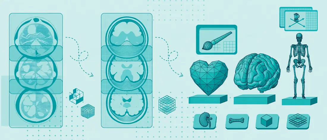
Modern workflows often begin with scan data. Specialists import DICOM files, segment organs and tissues, and convert them into 3D meshes. Then artists refine the surfaces, add materials, and light the scene for clarity.
This approach offers clear advantages:
- Patient-specific accuracy: Teams can model real anatomy instead of an idealized average.
- Measurable geometry: Surgeons can verify distances, angles, and spatial relationships.
- Reusable assets: One model can generate stills, animations, and interactive views.
Software That Changed the Workflow
Adobe Illustrator (1987) and Photoshop (1990) moved traditional techniques onto the screen. Later, sculpting and 3D tools such as ZBrush, Blender, Cinema 4D, and Maya expanded what was possible. Each tool serves a different purpose, from organic sculpting to physically based rendering. For a practical breakdown, see our comparison of digital sculpting and rendering tools used by medical artists.
3D, Animation, VR, and AR
Digital illustration now extends beyond the page. Studios create:
- Interactive 3D models for patient education
- Surgical animations for training and marketing
- Virtual and augmented reality simulations for medical education
- Regulatory-ready device visuals for submissions and sales teams
Understanding the difference between static art and motion helps teams choose the right format. Our article explaining where still illustration and motion graphics differ is a useful starting point.
AI and the Question of Accuracy
Generative AI has entered the conversation. It can speed up concept exploration, but it also invents anatomy that looks convincing and is wrong. For example, tools may add extra bones, distort organs, or mix up labels. Consequently, many publishers restrict AI-generated scientific images, and expert review is essential for clinical content.
The lesson from history is clear. Vesalius replaced inherited error with evidence. Modern teams must do the same by verifying every structure against reliable references.
Traditional vs Digital Medical Illustration: Comparison Table
Both approaches still have a place. This table helps you choose between them.
| Factor | Traditional (Ink, Carbon Dust, Watercolor) | Digital (2D and 3D) |
|---|---|---|
| Typical tools | Pen, carbon dust, gouache, airbrush | Tablet, vector software, 3D sculpting, rendering engines |
| Revision speed | Slow; changes often require redrawing | Fast; layers and models adjust easily |
| Data integration | Based on observation and reference | Can use CT, MRI, and segmentation data |
| Output formats | Print plates and scanned art | Print, web, video, AR, VR, and 3D files |
| Scalability | One original per image | Reusable assets and consistent series |
| Best for | Fine art style, historical reproduction | Journals, patient education, device marketing, training |
| Accuracy risk | Depends on artist reference | Depends on source data and expert review |
How a Modern Medical Illustration Is Made: Step-by-Step
Our studio workflow mirrors the Vesalius model of collaboration between expert and artist. Here is the process in six steps:

- Define the purpose and audience. Decide whether the image is for a journal, a patient handout, or a sales deck. Each use changes the level of detail.
- Gather reference material. Collect scans, specimens, surgical notes, and published atlases. Confirm that every source is reliable.
- Build a rough sketch or blockout. Share composition and labeling early so the expert can correct structure before polishing begins.
- Model, render, or paint the final art. Choose 2D or 3D based on the story the image must tell.
- Run a clinical accuracy review. Ask a physician or scientist to verify every structure, label, and color choice.
- Deliver in the right formats. Provide vector files, high-resolution raster files, and web-optimized versions. Our guide to AI and EPS vector file formats covers the details.
Practical Insights: What the History of Medical Illustration Teaches Today
Looking back at five centuries of work offers practical guidance for any team that commissions visuals.
- Accuracy builds trust. Vesalius earned lasting authority because his images matched reality. Likewise, today’s audiences trust brands that show correct anatomy.
- Clarity beats realism. Brödel’s best plates removed distractions. Therefore, your illustration should show what the viewer needs and nothing more.
- Collaboration produces better work. The strongest historical books paired experts with skilled artists. The same partnership improves modern projects.
- Format matters. Woodcut, lithograph, and 3D render each suit specific goals. Match the medium to the audience.
- Technology changes, principles stay. Observation, verification, and storytelling have guided the field since 1543.
Common Mistakes When Studying or Commissioning Medical Illustration
Many people repeat the same errors, whether they research the field or hire a studio. Avoid these pitfalls:
- Treating illustration as decoration. Beautiful images that carry errors can mislead students and damage credibility.
- Skipping expert review. Without a clinician check, even small label mistakes slip into print.
- Ignoring the audience. A surgeon needs different detail than a patient. Define the viewer first.
- Using AI output without verification. Generated anatomy often looks right at a glance and fails under scrutiny.
- Overlooking file requirements. Journals and printers demand specific resolutions, color modes, and formats. Review journal author guidelines for illustrations before you start.
- Confusing historical style with accuracy. Some vintage images look elegant but contain outdated or incorrect anatomy.
Expert Tips for Getting the Most From Medical Illustration
Professional illustrators share a few habits that raise quality on every project:
- Start with a clear brief. State the message, the audience, and the publication target in the first conversation.
- Share your best reference. Send scans, papers, and surgical notes early, because strong reference prevents rework.
- Review sketches, not just finals. Early feedback costs less than late changes.
- Limit labels and color. Fewer, clearer callouts improve comprehension.
- Plan for reuse. Ask for layered files and consistent styles so you can adapt visuals for papers, slides, and web pages.
- Check licensing. Confirm usage rights for print, digital, and promotional use.
FAQ: History of Medical Illustration
What is the history of medical illustration in simple terms?
The history of medical illustration describes how artists and clinicians created images to teach, record, and communicate human anatomy and disease. It moved from hand-drawn diagrams and woodcuts to engraving, lithography, photography, and now 3D digital rendering.
Who is the father of medical illustration?
Many historians credit Andreas Vesalius with launching modern anatomical illustration through his 1543 Fabrica. Others name Max Brödel the father of modern medical illustration because he created the first professional training program in 1911.
Why was Vesalius’s Fabrica so important?
Vesalius based his images on direct human dissection and corrected many errors in Galen’s teaching. Moreover, he combined text, art, and printing into one integrated system. That approach shaped anatomical education for centuries.
How did Max Brödel change medical illustration?
Brödel introduced the carbon dust technique, drew from the surgeon’s viewpoint, and founded the Department of Art as Applied to Medicine at Johns Hopkins in 1911. Consequently, medical illustration became a formal profession with clear training standards.
How has digital technology changed medical illustration?
Digital tools allow illustrators to work from CT and MRI data, build 3D models, and deliver interactive content. In addition, revisions happen faster, and teams can reuse one asset across print, web, video, and VR.
Is traditional medical illustration still used today?
Yes. Many illustrators still use pen, watercolor, and carbon dust for specific styles. However, most commercial projects now rely on digital workflows because they offer speed, flexibility, and multiple output formats.
Can AI replace medical illustrators?
Not safely. AI can support brainstorming, but it often produces inaccurate anatomy and unverifiable details. Therefore, clinical and scientific content still needs trained illustrators and expert review.
Conclusion: Why the History of Medical Illustration Still Matters
The history of medical illustration shows a consistent pattern. Each breakthrough, from Vesalius’s woodcuts to Brödel’s carbon dust plates to today’s 3D renders, made medical knowledge clearer and more trustworthy. Meanwhile, the tools changed, but the principles stayed the same: observe carefully, verify with experts, and communicate with purpose.
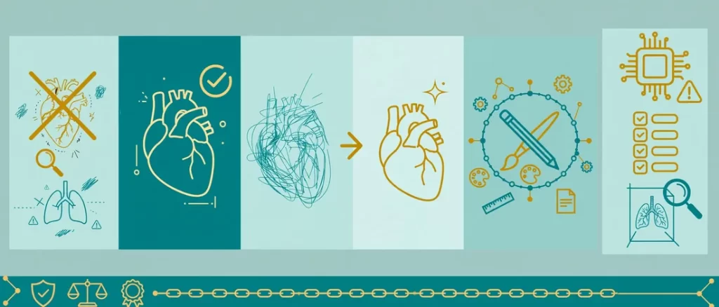
Key points to remember:
- Vesalius turned anatomy into an evidence-based visual science in 1543.
- Brödel built the profession and its training standards.
- Digital workflows now connect scan data, 3D modeling, and interactive media.
- Accuracy and expert review remain non-negotiable.
If you want visuals that carry that same standard of accuracy, commission custom medical illustrations that make your research, device, or training clear and credible. For more guidance, browse more articles in our medical illustration blog.