3D medical illustrations are scientifically accurate, three-dimensionally rendered visual assets produced using specialized CGI and anatomical modeling software to depict human anatomy, pathophysiology, medical devices, and biological processes with spatial depth, photorealistic rendering, and multi-axis viewing capability. Unlike traditional flat artwork, these illustrations are constructed from volumetric polygon meshes, texture-mapped surfaces, and physically based rendering (PBR) pipelines, enabling clinical-grade accuracy across applications ranging from pharmaceutical mechanism-of-action (MoA) visuals to surgical planning overlays. As demand for precise biomedical communication intensifies across healthcare, life sciences, and medical-legal markets, 3D medical illustrations have become the definitive standard for conveying complex anatomical and physiological concepts with unambiguous visual clarity.
The global medical animation and illustration market was valued at over USD 511 million in 2025 and is projected to reach nearly USD 2.6 billion by 2034, growing at a compound annual growth rate (CAGR) of nearly 18% (Precedence Research, 2025). That trajectory reflects a fundamental truth: when science must communicate, it must visualize – and three-dimensional rendering is the most powerful tool available today.
Whether you are a pharmaceutical brand manager, a medical device manufacturer, a healthcare educator, or a litigation attorney, understanding what 3D medical illustration offers – and how it differs from other visual formats – is critical to choosing the right communication strategy for your audience.
Table of Contents
What is 3D Medical Illustration?
At its core, a 3D medical illustration is a digitally constructed, scientifically grounded visual representation of biological structures, medical devices, or clinical procedures. Unlike photography or 2D line art, these illustrations exist as fully navigable three-dimensional objects within a digital environment. They can be rotated, sliced, lit from any angle, and rendered at any resolution – before a single final image is exported.
The field sits at the intersection of biomedical science, fine art, and computational visualization technology. Practitioners who create these visuals – known as medical illustrators – typically hold advanced degrees in medical illustration or biomedical communications. They combine an understanding of gross anatomy, histology, and cellular biology with proficiency in software tools such as Autodesk Maya, ZBrush, Cinema 4D, and Blender, alongside anatomical reference sources including DICOM imaging data from CT and MRI scans.
What makes a 3D medical illustration “medical”?
The defining distinction is scientific rigor. A high-quality 3D medical illustration must:
- Accurately represent anatomical proportions, tissue layers, and spatial relationships
- Reflect the current, peer-reviewed understanding of the biological process being depicted
- Be created or reviewed by certified medical illustrators with domain expertise
- Meet the accuracy standards required by the intended audience – whether clinicians, regulators, patients, or jurors
This combination of artistic skill and scientific accountability is what separates professional 3D medical illustration from general-purpose 3D rendering.
3D vs. 2D Medical Illustration: Key Differences
To fully appreciate the value of three-dimensional rendering, it helps to understand where it diverges from traditional 2D approaches. Both have legitimate roles in biomedical communication – but they serve different needs.
For a broader comparison of how illustration fits within visual medical communication, read our overview of what medical illustration is and how it works.
Comparison Table: 3D vs. 2D Medical Illustration
| Feature | 2D Medical Illustration | 3D Medical Illustration |
|---|---|---|
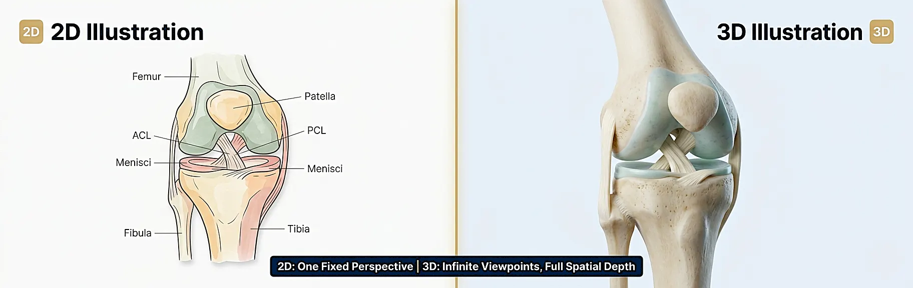
| Spatial Depth | Simulated through shading and perspective | Fully volumetric; true spatial depth |
| Viewing Angles | Fixed at creation; one perspective per image | Unlimited viewing angles from a single model |
| Reusability | Each new angle requires new artwork | Single model can generate hundreds of outputs |
| Photorealism | Limited; stylized rendering | High; physically based rendering (PBR) |
| Animation Capability | Not applicable | Directly animated from the same 3D asset |
| Production Time | Faster for single-image outputs | Longer initial build; faster for multi-view outputs |
| Cost Efficiency | Lower for one-off images | Higher ROI across multi-format campaigns |
| Regulatory Submissions | Widely accepted | Increasingly preferred for device marketing |
| Patient Comprehension | Good for simple concepts | Significantly higher for complex spatial anatomy (Frontiers in Pediatrics, 2021) |
| Surgical Planning | Limited utility | Used directly in pre-operative visualization |
As research published in Frontiers in Pediatrics confirms, three-dimensional visualization modalities consistently outperform two-dimensional images when students and patients need to understand spatial anatomical relationships. That finding has significant implications for any organization using visuals to educate, train, or persuade.
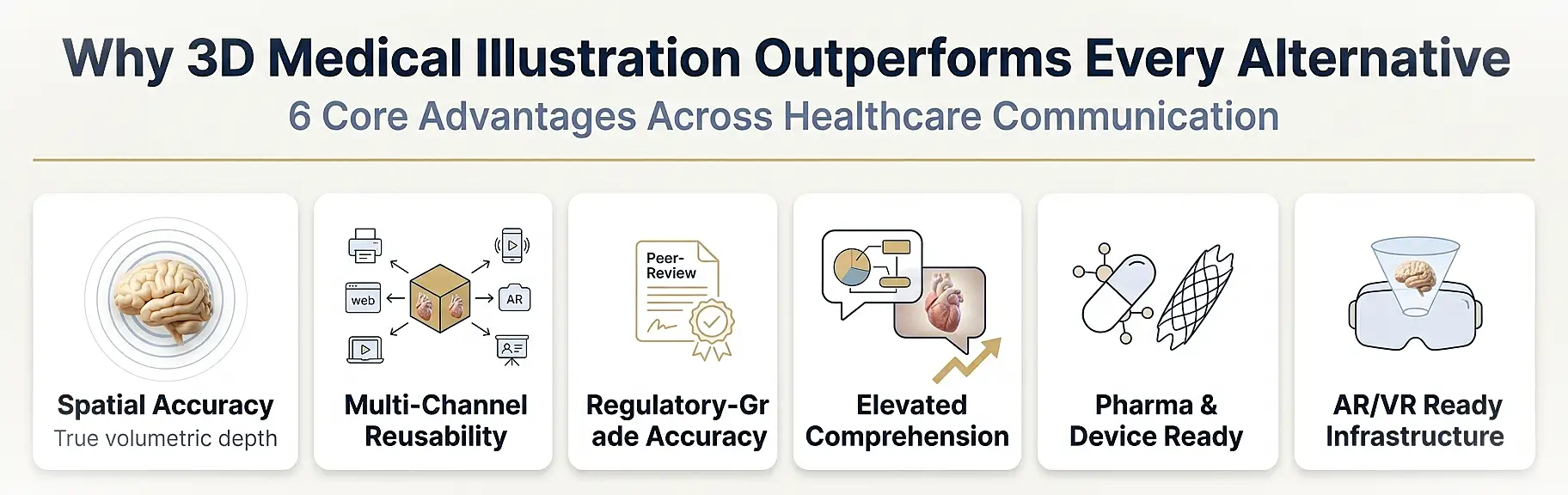
Core Benefits of 3D Medical Illustrations
1. Unmatched Spatial Accuracy and Depth
The human body is a three-dimensional structure. Representing it in three dimensions is not simply an aesthetic preference – it is the most scientifically honest approach. 3D medical illustrations allow the viewer to perceive depth, layering, and spatial relationships between structures in ways that flat artwork cannot replicate. A surgeon reviewing a 3D model of a patient’s vascular anatomy before an intervention gains far more actionable spatial understanding than one working from a 2D diagram.
2. Versatility Across Multiple Deliverables
One of the most compelling practical advantages of 3D illustration is asset reusability. A single, rigorously built 3D model of a spinal implant, for example, can generate:
- Still images for journal submissions and regulatory filings
- Turntable renders for trade show displays
- Exploded-view diagrams for surgical training manuals
- Interactive web embeds for HCP portals
- Animation sequences for patient education videos
- Augmented reality overlays for device demonstration
This multi-output capability means that the initial investment in a 3D asset pays dividends across every communication channel simultaneously.
3. Enhanced Comprehension for Complex Concepts
Mechanisms of disease, drug-receptor binding interactions, cellular signaling pathways, and multi-step surgical procedures are all notoriously difficult to explain through text or 2D imagery alone. Three-dimensional rendering breaks these complex processes into visually navigable sequences. Viewers can follow a pharmacological agent through a receptor cascade, watch a stent deployment unfold layer by layer, or observe how a viral particle interacts with a host cell membrane – all with the spatial clarity that drives genuine understanding.
4. Regulatory and Clinical Credibility
In pharmaceutical marketing and medical device promotion, accuracy is not optional – it is legally mandated. 3D medical illustrations produced by certified, board-eligible medical illustrators carry scientific credibility that generic stock imagery cannot provide. They can be submitted as part of FDA marketing authorization packages, used in peer-reviewed publications, and presented in legal proceedings without compromising the accuracy standards those contexts demand.
5. Scalability for Large Campaigns
Pharmaceutical companies, medical device manufacturers, and healthcare systems frequently require visual assets at scale – dozens of procedure illustrations, hundreds of product views, or complete anatomical libraries. Because 3D workflows are modular (individual anatomical components can be combined, isolated, and reconfigured), scaling a 3D-based visual program is far more efficient than commissioning dozens of separate 2D artworks.
6. Future-Proof Asset Investment
3D models built today can be repurposed for emerging delivery formats – virtual reality surgical simulators, augmented reality anatomy apps, interactive digital patient education platforms, and AI-driven clinical decision tools. Organizations that invest in high-quality 3D medical illustration assets are building a visual infrastructure that remains relevant as technology evolves.
.
Primary Use Cases and Industry Applications
3D medical illustrations serve a remarkably broad range of professional contexts. Here is a structured overview of the most significant application areas:
Pharmaceutical and Biotech Marketing
Pharmaceutical brands use 3D medical illustrations to visualize mechanism-of-action (MoA) sequences, drug-target interactions, and clinical outcome concepts. These assets appear in detail aids used by medical sales representatives, in HCP-targeted digital campaigns, and in direct-to-consumer advertising where visual clarity about how a drug works builds prescriber and patient confidence.
Custom imagery developed for these purposes must pass through rigorous medical-legal review (MLR) processes. Certified medical illustrators understand these workflows and build accuracy documentation into the production process from the outset.
Medical Device Marketing and Training
Medical device manufacturers rely heavily on 3D illustration for product visualization. Whether demonstrating how an orthopedic implant integrates with bone architecture, showing the deployment mechanism of an endovascular stent, or illustrating the optical pathway through a surgical microscope, 3D rendering communicates device function with a precision that product photography alone cannot achieve.
These visuals support:
- Pre-sales presentations to surgeons and hospital procurement teams
- IFU (Instructions for Use) documentation
- Distributor and sales team training materials
- Investor relations and fundraising decks
For a detailed look at how illustration serves device marketing specifically, our article on medical device and product illustration covers the subject in depth.
Medical Education and Training
Medical schools, nursing programs, allied health training institutions, and continuing medical education (CME) providers all use 3D medical illustrations to teach anatomy, physiology, pathology, and clinical procedures. Research consistently demonstrates that 3D visualization modalities improve spatial comprehension of anatomical structures compared to traditional 2D atlas materials (NIH/PMC, 2021).
Applications include:
- Surgical technique atlases
- Anatomy curriculum resources
- Pathophysiology explainers for nursing and allied health students
- CME-accredited visual learning modules
- Simulation training materials
Patient Education and Informed Consent
Communicating a diagnosis, proposed surgery, or chronic disease management plan to a patient requires visuals that are accurate, clear, and emotionally accessible. 3D medical illustrations occupy a valuable middle ground: they are scientifically precise enough to satisfy clinical standards, yet visually approachable enough for a patient with no medical background.
Common patient education applications include pre-operative procedure explanations, post-diagnosis condition overviews, medication mechanism explainers, and rehabilitation exercise guides. When patients genuinely understand what is happening inside their bodies, they make more informed decisions and demonstrate better treatment adherence.
Medical-Legal and Litigation Support
In personal injury, medical malpractice, and product liability cases, 3D medical illustrations function as demonstrative evidence. Attorneys use them to explain injuries, surgical errors, anatomical vulnerabilities, and causation mechanisms to judges and juries who have no medical training.
The accuracy standards for litigation-support illustration are exceptionally high. Every structural detail must be defensible by the testifying medical expert, and the illustration must not misrepresent anatomy or amplify injury beyond what the clinical record supports. Certified medical illustrators who specialize in legal work understand these constraints.
Scientific Publication and Research Communication
Journals, research institutions, and academic publishers commission 3D medical illustrations for figures in peer-reviewed articles, book chapters, grant applications, and conference presentations. The journal submission process for scientific illustrations follows specific author guidelines; our guide to author guidelines for illustrations in medical and scientific journals outlines exactly what publishers expect.
Digital Health, AR, and VR Applications
Emerging digital health platforms are driving a new category of demand for 3D medical assets. Augmented reality surgical guidance tools, VR anatomy training platforms, and interactive patient education applications all require 3D models with the polygon density, texture fidelity, and rigging capability to perform in real-time rendering environments.
Real-World Examples of 3D Medical Illustration
Understanding the theory is valuable. Seeing how 3D medical illustration functions in practice makes the value proposition concrete. Below are five representative examples spanning the major application categories:
Example 1: Cardiac Anatomy for HCP Digital Campaign
A cardiovascular pharmaceutical company required a fully articulated 3D model of the left ventricle to illustrate the mechanism of action of a new heart failure medication. The illustration depicted ion channel behavior at the myocardial cell membrane level, with a progressive zoom sequence moving from whole-heart anatomy to cellular ultrastructure. The same model generated 12 distinct still frames, three animated sequences, and a downloadable PDF leave-behind.
Example 2: Spinal Implant System for Surgical Training
An orthopedic device manufacturer commissioned a complete 3D illustration library for a posterior lumbar interbody fusion (PLIF) system. The library included exploded-view cage positioning diagrams, vertebral cross-sections showing bone-implant integration, screw trajectory illustrations, and a step-by-step surgical technique sequence. The entire library was built from a single master 3D model set, ensuring anatomical consistency across all outputs.
Example 3: Oncology Mechanism-of-Disease for Patient Education
An oncology center needed patient-facing materials explaining how colorectal cancer develops from normal mucosal epithelium through adenoma to carcinoma. The 3D illustration series used accessible rendering styles – retaining scientific accuracy while avoiding the clinical detachment of photorealistic renders. Patient comprehension scores improved significantly versus the previous text-and-diagram approach.
Example 4: Knee Injury for Medical-Legal Use
A personal injury attorney required 3D medical illustrations showing the anatomical structures involved in a complex ligamentous knee injury. The illustrations depicted the ACL, PCL, medial meniscus, and associated soft tissue in their normal state, followed by a second illustration set showing the injury pattern consistent with the client’s MRI findings. The images were used as jury exhibits during trial, presented by the testifying orthopedic surgeon.
Example 5: Surgical Robotics Device for Investor Deck
A surgical robotics startup needed visualization assets for a Series B fundraising presentation. The 3D illustrations depicted the robotic arm’s operative envelope within the abdominal cavity, instrument tip articulation, and tissue interaction – communicating technical capability to non-clinical investors with no surgical background.
The Creation Process: How 3D Medical Illustrations Are Made
Professional 3D medical illustration follows a structured production pipeline. Understanding this process helps clients set realistic expectations, brief their creative partners accurately, and avoid costly revision cycles.
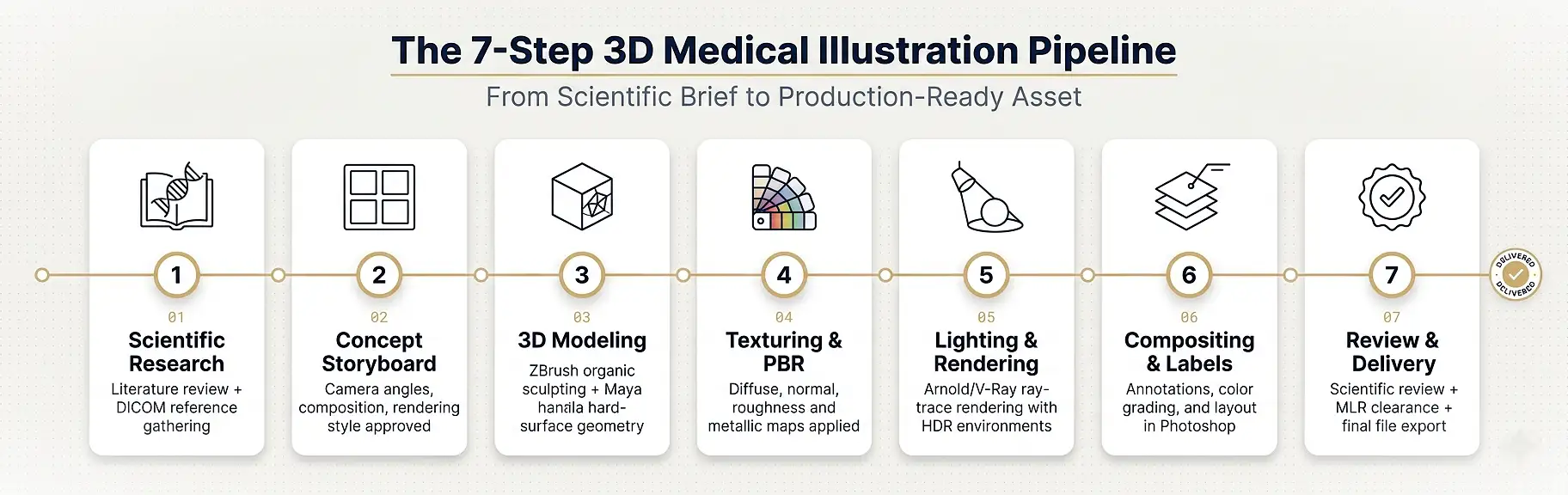
Step 1: Scientific Research and Reference Gathering
Before any geometry is created, the medical illustrator conducts thorough scientific research. This phase involves:
- Reviewing peer-reviewed anatomical literature and pathology references
- Consulting DICOM imaging data (CT, MRI scans) where available
- Reviewing product documentation for device illustrations
- Clarifying the scientific brief with subject matter experts or client medical advisors
For an overview of the software tools used at this stage and throughout production, see our dedicated article on 3D medical illustration software and platforms.
Step 2: Concept Development and Storyboarding
For multi-image series or sequences, the illustrator develops a visual storyboard – a layout showing which views, cross-sections, and callouts are needed. This stage establishes camera angles, composition, color palette, and rendering style (photorealistic, stylized, diagrammatic) before any time-intensive modeling begins.
Step 3: 3D Modeling and Geometry Construction
The core production phase involves building polygon meshes that accurately represent the anatomical structures or devices. Illustrators use tools such as ZBrush for organic tissue sculpting, Autodesk Maya or Cinema 4D for hard-surface device geometry, and 3D Slicer for segmenting real patient DICOM data into usable mesh references.
Key modeling considerations include:
- Topological accuracy relative to reference anatomy
- Polygon density appropriate for the intended output (print, screen, real-time AR)
- Correct spatial layering of tissue planes (skin, fascia, muscle, bone, vasculature)
- Anatomical landmark accuracy for structures that will be labeled or called out
Step 4: Texturing and Surface Properties
Once the geometry is approved, surfaces are textured to convey material properties. Skin has subsurface scattering. Bone has cortical density variation. Surgical instruments have metallic specularity. These surface properties are applied using physically based rendering (PBR) texture maps – including diffuse, normal, roughness, and metallic channels – that behave accurately under any lighting condition.
Step 5: Lighting and Rendering
Lighting design is one of the most consequential decisions in 3D medical illustration. The wrong light placement can obscure anatomical detail, create misleading shadows, or make a structure appear disconnected from its environment. Medical illustrators use HDR lighting environments, area lights, and carefully placed rim lights to reveal form while maintaining the visual hierarchy that guides the viewer’s eye to the clinically important structures.
Rendering is typically performed using ray-trace or path-trace engines such as Arnold, V-Ray, or Cycles, producing final images with accurate light behavior.
Step 6: Post-Processing and Annotations
Final rendered images are composited in Adobe Photoshop or After Effects, where labels, leader lines, color grading, and any stylistic finishing are applied. For publication submissions, image specifications follow journal guidelines. For pharmaceutical materials, the output goes through MLR review before distribution.
Step 7: Review, Revision, and Final Delivery
The completed illustrations are reviewed against the scientific brief, with input from the client’s medical and regulatory stakeholders. Revisions address any anatomical inaccuracies, compositional issues, or labeling corrections before final file delivery.
Common Mistakes to Avoid When Commissioning 3D Medical Illustrations
Even organizations with significant experience in healthcare marketing frequently make avoidable errors when commissioning 3D medical illustration. Here are the most consequential ones:
1. Prioritizing aesthetics over accuracy A visually stunning illustration that misrepresents anatomy creates legal and regulatory liability, particularly in pharmaceutical promotion and litigation contexts. Always require demonstrable scientific review as part of the production process.
2. Underestimating the scientific brief Vague briefs produce vague illustrations. The more specific and scientifically detailed your initial brief – including the exact anatomical structures, clinical context, audience, and intended use – the more accurately the illustrator can match your needs in fewer revision cycles.
3. Choosing generalist 3D studios over medical specialists General-purpose 3D visualization studios produce technically excellent renders, but they lack the biomedical training to ensure anatomical accuracy. For any application where clinical credibility matters, work exclusively with certified or board-eligible medical illustrators.
4. Ignoring deliverable format requirements early A model built for print output may not have the optimized polygon density for real-time AR rendering. Specify all intended output formats – print, digital, animated, interactive – at the outset of the project.
5. Skipping the storyboard phase Jumping straight to modeling without approving a visual storyboard is one of the most common causes of expensive late-stage revisions. Approve the concept before the high-investment production work begins.
6. Not securing usage rights Ensure your contract clearly specifies who retains ownership of the 3D model assets (not just the final images), and what usage rights are granted for various distribution channels and durations.
Expert Tips for Getting the Most from Your 3D Medical Visuals
Drawing from best practices in biomedical communication and visual design, here are actionable recommendations for maximizing the impact of your 3D medical illustration investments:
- Plan for multi-channel output from day one. Commission models with all intended applications in mind so the 3D assets can serve print, digital, animated, and interactive formats without rebuilding.
- Involve your medical and regulatory teams early. The earlier clinical accuracy and regulatory language requirements are established, the smoother the MLR review process will be.
- Use cross-sectional views deliberately. Some of the most powerful 3D medical illustrations are cutaway cross-sections that reveal internal structures invisible in surface-level renders. Plan these into your storyboard.
- Build an anatomical asset library. For organizations with recurring illustration needs, commissioning a modular asset library (core anatomical structures, signature device components) delivers dramatically better long-term ROI than one-off commissions.
- Align rendering style with your audience. Photorealistic renders work best for HCP and scientific audiences; more stylized, diagrammatic rendering with clear labels often communicates more effectively to patients and non-clinical decision makers.
- Ensure ALT text and accessibility for all digital uses. Every 3D medical illustration used in digital contexts should have descriptive ALT text that conveys the medical content of the image for screen reader accessibility.
For a broader look at why visual communication is central to effective healthcare practice, our article on the importance of medical illustrations across the healthcare industry provides an in-depth perspective worth reviewing.
Choosing the Right 3D Medical Illustration Partner
Not all 3D visualization providers are equipped to meet the standards required in medical, pharmaceutical, and clinical communication contexts. When evaluating potential partners, consider the following criteria:
Credentials and Training
Look for illustrators who hold a Master of Science in Medical Illustration from an accredited program (such as those affiliated with the Association of Medical Illustrators) or who are certified by the Board of Certification of Medical Illustrators (BCMI). These credentials demonstrate that the illustrator has undergone rigorous biomedical science training alongside their technical skills.
Portfolio Depth and Domain Expertise
Review portfolio work for anatomical accuracy, rendering quality, and experience in your specific application area – pharmaceutical, device, legal, educational. A studio with deep experience in cardiovascular device illustration may or may not be the strongest choice for a neurological patient education project.
Scientific Review Process
Ask specifically how scientific accuracy is verified. Leading studios build in subject matter expert review, literature citation documentation, and client medical advisory review as standard process steps – not optional add-ons.
Software and Technology Capability
For projects requiring AR, VR, or interactive delivery, confirm that the studio’s pipeline includes real-time engine capability (Unreal Engine, Unity) alongside traditional rendering software.
Client References and Case Studies
Request case studies from comparable projects and speak directly with previous clients about production timeline adherence, revision responsiveness, and scientific accuracy standards.
FAQ
What is a 3D medical illustration?
A 3D medical illustration is a scientifically accurate, three-dimensionally rendered digital artwork depicting human anatomy, biological processes, medical devices, or clinical procedures. It is created using CGI software by trained medical illustrators who combine biomedical science knowledge with advanced visualization skills. Unlike 2D illustrations, 3D medical illustrations can be viewed from multiple angles, animated, and deployed across print, digital, and interactive formats.
How are 3D medical illustrations different from medical animation?
3D medical illustrations produce still rendered images from three-dimensional models. Medical animations use those same 3D models to produce motion sequences showing biological processes, device deployments, or procedural steps unfolding over time. Both formats share the same 3D asset infrastructure; the key difference is static versus dynamic output. For a detailed breakdown of these two formats, see our comparison of medical illustration versus medical animation.
What software is used to create 3D medical illustrations?
The most widely used tools include Autodesk Maya, ZBrush, Cinema 4D, Blender, and 3ds Max for modeling; Arnold, V-Ray, KeyShot, and Cycles for rendering; and Adobe Photoshop and After Effects for compositing and annotation. DICOM processing tools such as 3D Slicer are used to convert CT and MRI data into usable mesh references.
How much do 3D medical illustrations cost?
Pricing varies significantly based on anatomical complexity, level of photorealism, number of deliverable outputs, and timeline. A single-view still illustration of a medical device typically starts at several hundred dollars; a complex multi-view anatomical series or complete mechanism-of-action sequence for pharmaceutical use can range into tens of thousands. Organizations with recurring needs benefit from retainer or library-build arrangements that deliver significantly better per-asset economics.
How long does it take to create a 3D medical illustration?
Production timelines depend on project complexity. A single anatomical still can be delivered in one to two weeks. A multi-scene pharmaceutical MoA sequence with full scientific review may require six to twelve weeks. Rush timelines are possible but typically carry a premium and require client responsiveness at review stages to maintain momentum.
Can 3D medical illustrations be used for FDA submissions?
Yes. 3D medical illustrations and diagrams produced to appropriate accuracy standards are routinely included in regulatory submissions, including device premarket notification (510(k)) packages and pharmaceutical marketing authorization applications. The key requirement is that all images accurately represent the device, drug, or anatomy as described in the scientific and clinical record.
What industries use 3D medical illustrations most?
The primary industries are pharmaceutical and biotech, medical device manufacturing, healthcare education and training, medical-legal and litigation support, scientific publishing, and digital health technology. Each has specific accuracy, style, and format requirements that a qualified medical illustration partner can address.
Conclusion
3D medical illustrations represent the most powerful tool available for communicating complex biomedical information with spatial accuracy, clinical credibility, and cross-channel versatility. From pharmaceutical mechanism-of-action campaigns to surgical training libraries, patient education materials to litigation demonstratives, the applications are as broad as the life sciences themselves – and the demand for high-quality 3D medical visualization is accelerating rapidly.
Organizations that invest in professionally produced 3D medical illustration assets gain a durable competitive advantage: visuals that are scientifically defensible, adaptable across every communication channel, and compelling enough to drive genuine audience understanding and engagement.
The difference between adequate and exceptional medical visualization is the depth of scientific expertise built into every polygon, texture, and light placement decision. That expertise is not incidental – it is the entire foundation of the discipline.
If your organization is ready to elevate its medical communication strategy with production-ready, scientifically accurate 3D visual assets, the team at The Medical Illustration Company is equipped to deliver. Commission custom 3D medical illustrations that meet your clinical, regulatory, and marketing standards – and experience the difference that specialist expertise makes.